Enhanced ultrasound tongue imaging
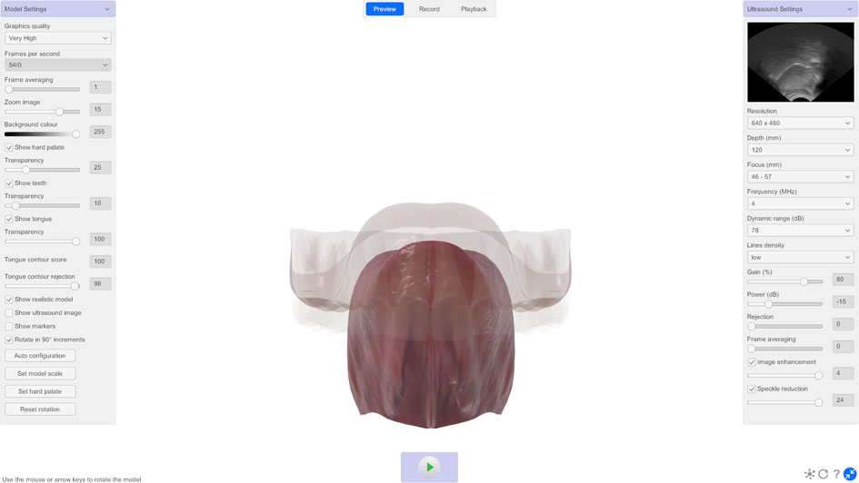
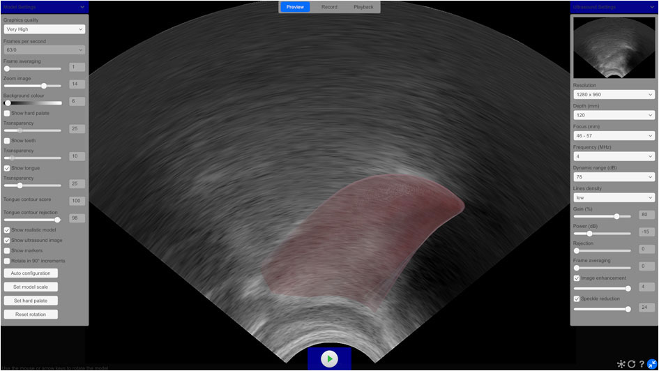
Conventional ultrasound tongue imaging is powerful, but the speckled greyscale images can sometimes be difficult to interpret, even for experienced users. LinguaSound 3D uses deep learning to estimate tongue surface contours from live ultrasound images and animates a rotatable 3D model of the tongue, hard palate and teeth in real time. It also provides tongue contour overlays, probe alignment feedback and an interactive biofeedback game with adjustable targets. Alongside these visual and biofeedback tools, timestamped tongue contour data can be exported for research and speech technology development. Together, these capabilities offer a clearer, more versatile way to explore tongue movement during speech.
How LinguaSound 3D works
Ultrasound tongue imaging (UTI) is a non-invasive technique used to view the shape, position and movements of the tongue during speech using high-frequency sound waves. A layer of acoustic gel is applied to an ultrasound probe placed beneath the chin. The probe emits sound waves that travel through the tissues and are reflected back when they encounter different structures within the tongue. The ultrasound scanner analyses these reflections to generate dynamic images of tongue shape and motion, which are transferred to the host computer via USB.
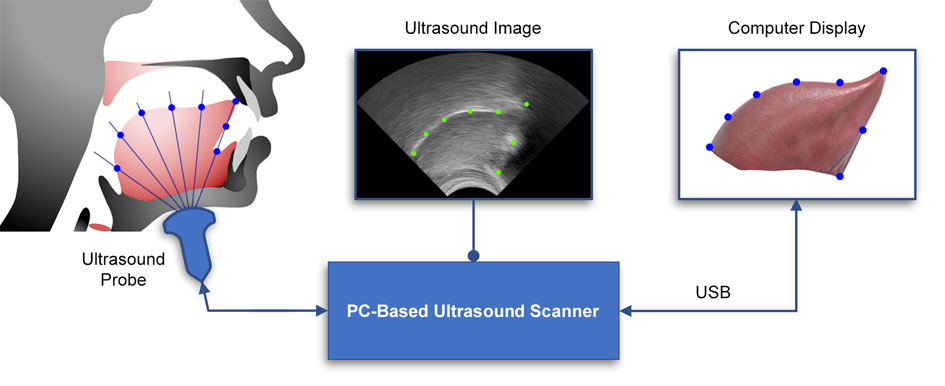
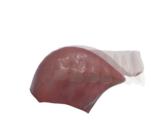
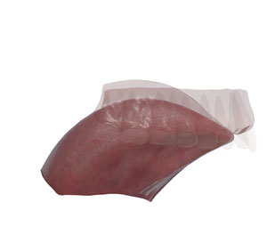
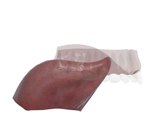
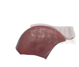
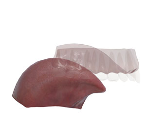
LinguaSound 3D uses a deep learning model to generate tongue contour estimations from the ultrasound images. The model was trained on thousands of hand-labelled midsagittal ultrasound images of both children and adults, using the DeepLabCut framework. These contours are then used to drive an animated 3D model of the tongue, hard palate and teeth in real time. The 3D model represents tongue position and movement based on the estimated contour, rather than a direct image of the speaker's tongue.
Hard palate estimation
Due to the air above the tongue surface reflecting sound waves back to the probe, conventional ultrasound tongue imaging cannot directly capture the hard palate. LinguaSound 3D estimates its position and size by detecting the maximum extent of the tongue contour during contact. Hard palate estimation is performed as a brief setup sequence at the start of each session and can be repeated to refine the position as required. For best results, the speaker should produce sounds that bring the tongue into contact with the alveolar, palatal and velar regions. This procedure is illustrated in the following video:
Probe alignment
Maintaining a consistent probe position is one of the challenges of ultrasound tongue imaging. Even small changes in probe orientation or pressure can alter the appearance of the tongue, making it difficult to compare recordings over time.
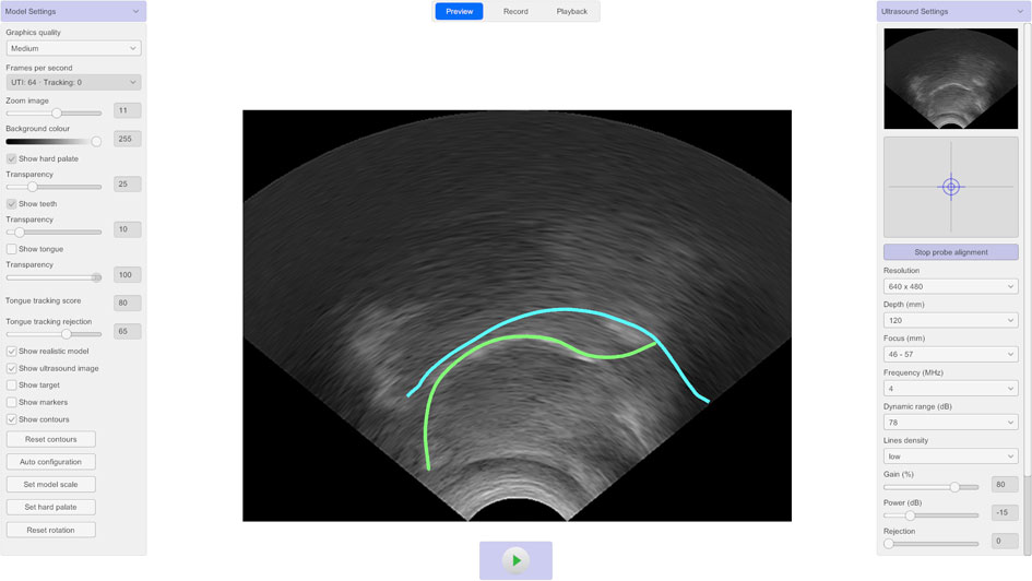
LinguaSound 3D includes a probe alignment tool that helps users maintain a consistent probe position throughout a session. Once a reference position has been set, a visual target indicator provides real-time feedback on changes in probe orientation and pressure. If the probe moves, the indicator shows both the direction and magnitude of the movement, allowing the original position to be restored quickly.
The Probe alignment tool can be used in Preview, Record and Playback modes, making it easy to monitor probe position during real-time biofeedback, data recording and playback of recorded sessions.
Tongue contour overlay
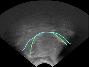
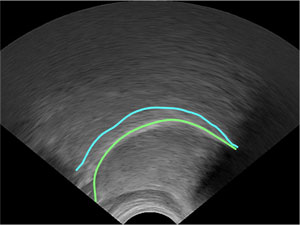
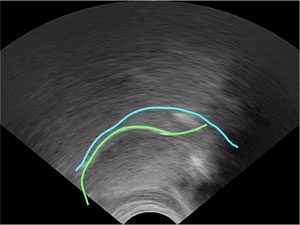
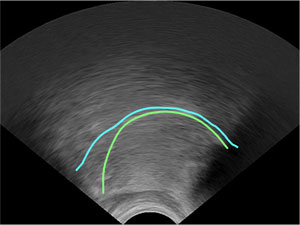
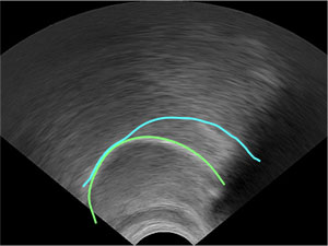
While the 3D model provides an intuitive view of tongue position and movement, some clinicians and researchers may prefer to work directly with the ultrasound image. LinguaSound 3D can display the estimated tongue contour as an overlay on the ultrasound image, allowing the tongue shape to be followed as it moves. The overlay is available in Preview, Record and Playback modes.
An accumulated contour can also be displayed, showing the maximum extent reached by the tongue as the contour is traced. Because the tongue cannot extend beyond the hard palate, this accumulated contour can be used to trace the shape of the palate as the speaker produces a range of sounds.
Both overlays are displayed as lightweight lines rather than a shaded reconstruction, keeping the underlying ultrasound image clearly visible. The tongue contour overlay can also be shown or hidden independently of the 3D model, giving users the flexibility to choose the view that best suits their needs.
Interactive game
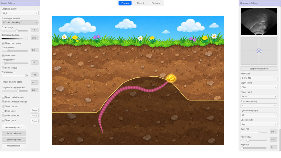
LinguaSound 3D includes an interactive game that presents real-time tongue movement as engaging visual biofeedback. The estimated tongue contour is represented by an animated worm, while the accumulated contour gradually forms the upper boundary of its cave — providing a visual approximation of the hard palate during speech. The game is available in Preview, Record and Playback modes.
A movable target in the form of a gold nugget can be positioned at a chosen location within the cave. When any part of the tongue contour touches the target, the gold nugget illuminates, providing an immediate visual reward.
The game provides an engaging way to practise and reinforce articulatory placement, with the movable target allowing different tongue positions to be explored. The game-based format can also make ultrasound tongue imaging feel more approachable and engaging for younger speakers, helping to maintain attention and motivation during use.
Tongue contour data export
Tongue movement provides a rich source of articulatory information for speech research and human-computer interaction. LinguaSound 3D converts live ultrasound tongue images into structured tongue contour data in real time, for use in articulatory research, silent speech interfaces, multimodal experiments, machine learning and other data-driven applications.
Each time a new tongue contour is estimated, LinguaSound 3D identifies eleven landmarks along the tongue surface, together with the hyoid bone, base of the mandible and mental spine. Each landmark is exported with its x and y coordinates and a confidence score. The results can be written continuously to CSV alongside the corresponding ultrasound frames.
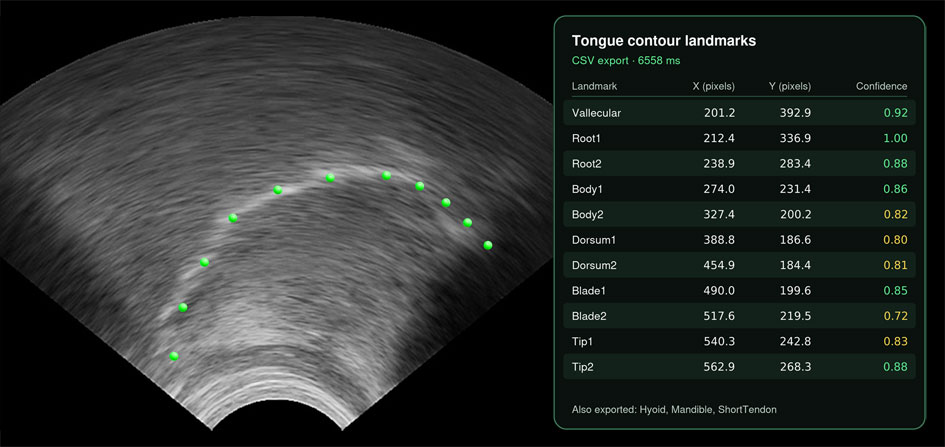
Each exported row includes a millisecond timestamp, allowing tongue movement to be aligned with audio, EMG, motion capture and other sensor data for multimodal analysis.
If you are developing speech technology, assistive devices, tongue-controlled interaction or research projects that need tongue movement data, please contact us to discuss your requirements.
Comparing conventional ultrasound tongue imaging with LinguaSound 3D
LinguaSound 3D makes conventional ultrasound tongue imaging easier to interpret. A comparison of the imaging techniques is illustrated below for a typical English speaker:
| Conventional UTI | Tongue contour overlays | 3D tongue render | |
|---|---|---|---|
| Velar Stop /k/ | 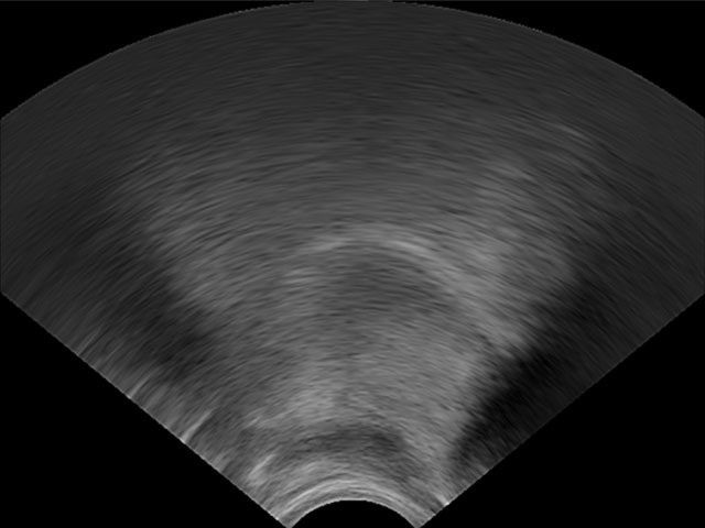 |
 |
 |
| Alveolar Stop /d/ | 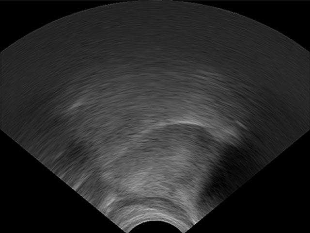 |
 |
 |
| Retroflex /r/ | 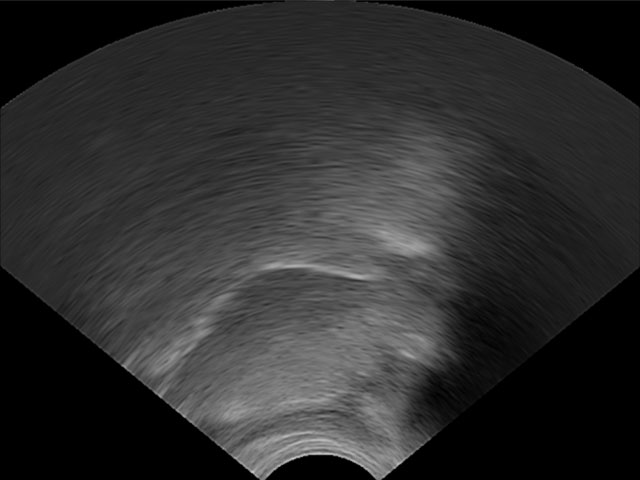 |
 |
 |
| High-Front Vowel /i/ | 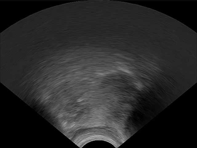 |
 |
 |
| Low-Back Vowel /a/ | 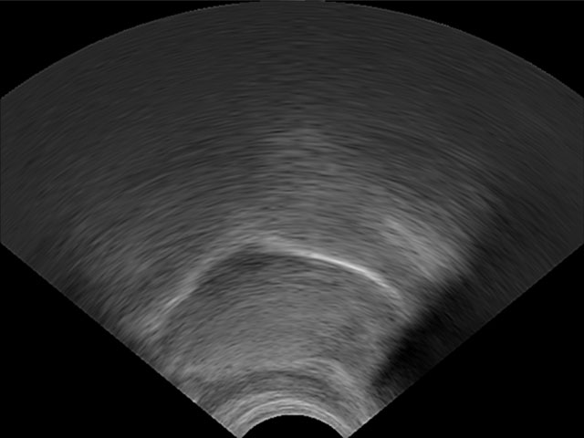 |
 |
 |
Compatible hardware
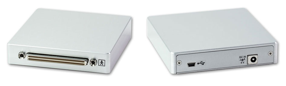
LinguaSound 3D is currently compatible with the Telemed MicrUs EXT-1H scanner and the MC4-2R20S-3 and MC10-5R10S-3 probes. The scanner connects to any compatible Windows laptop or desktop via USB. Compact, lightweight and fanless, it is suitable for use in clinical, classroom and laboratory settings. Contact us to discuss compatibility with your existing equipment or for purchasing guidance.
| Model | Type | Frequency Range | Radius | Scan Depth | Field of View | |
|---|---|---|---|---|---|---|
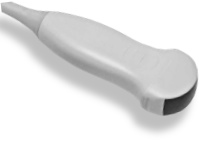 |
MC4-2R20S-3 | Convex | 2.0 - 4.0 MHz | 20 mm | 60 - 230 mm | 104° |
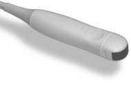 |
MC10-5R10S-3 | Micro Convex | 5.0 - 10.0 MHz | 10 mm | 30 - 150 mm | 156° |
LinguaSound 3D software
LinguaSound 3D is a Windows 64-bit application that combines real-time ultrasound imaging, tongue contour estimation and 3D visualisation in a single user interface.
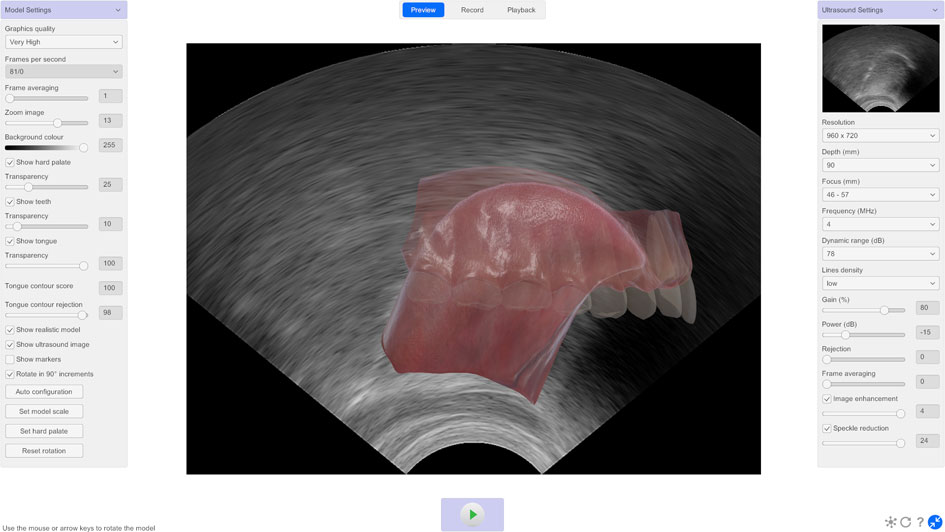
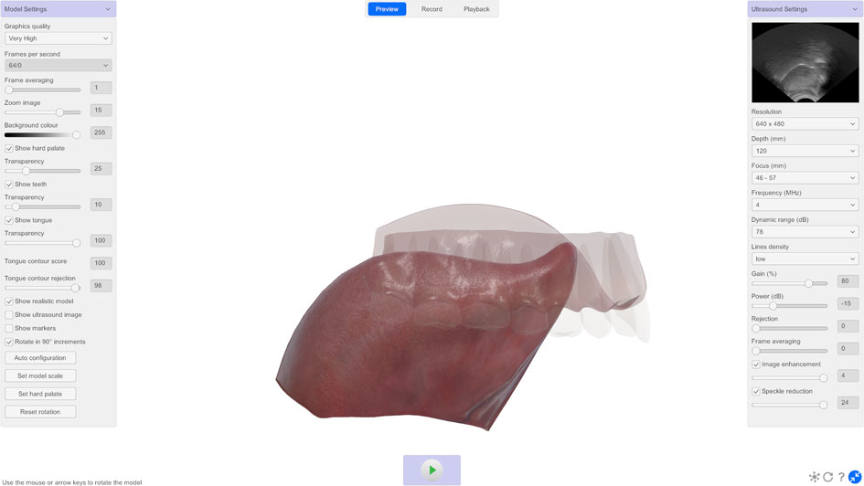
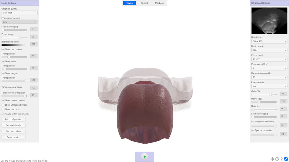
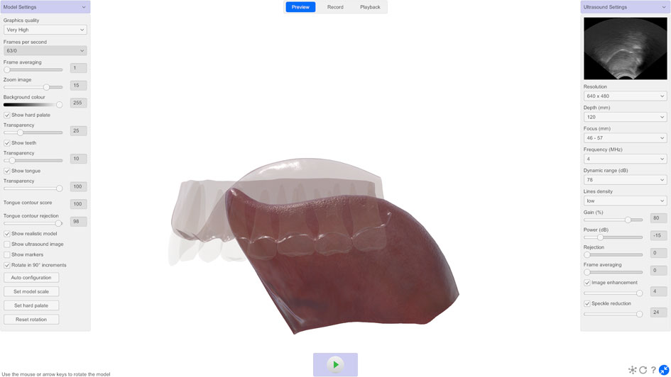
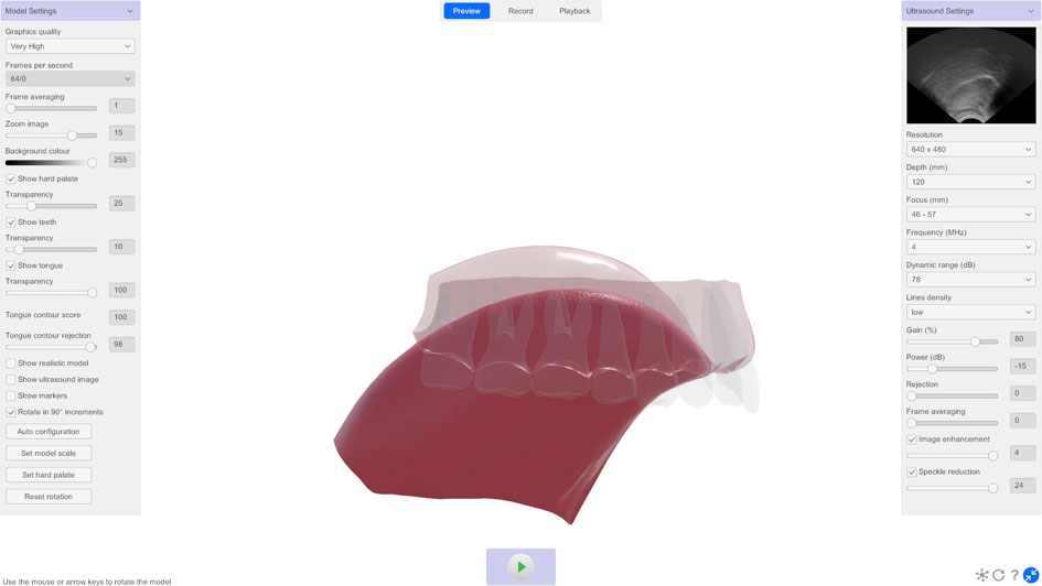
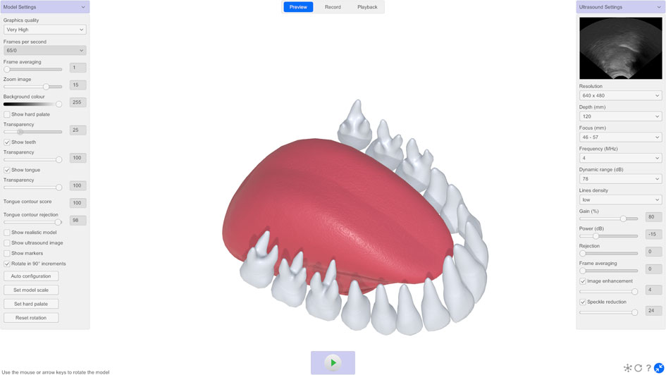
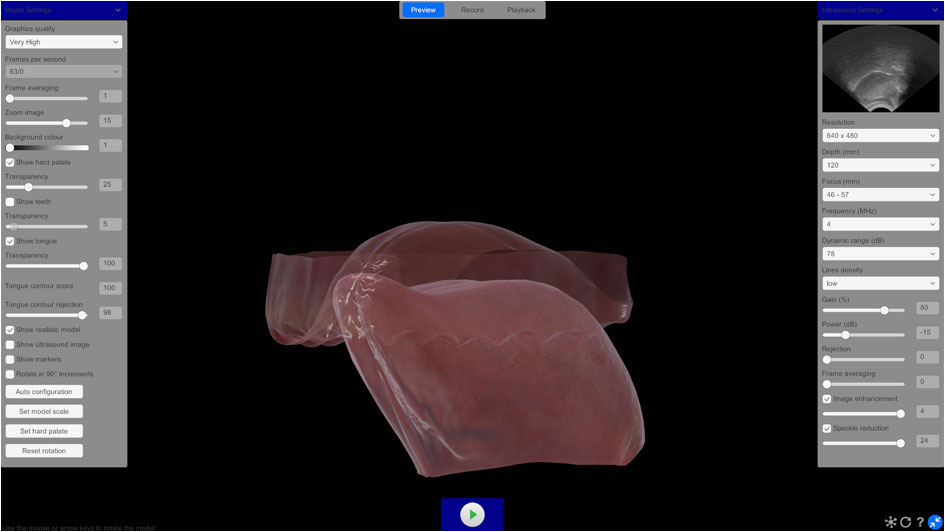
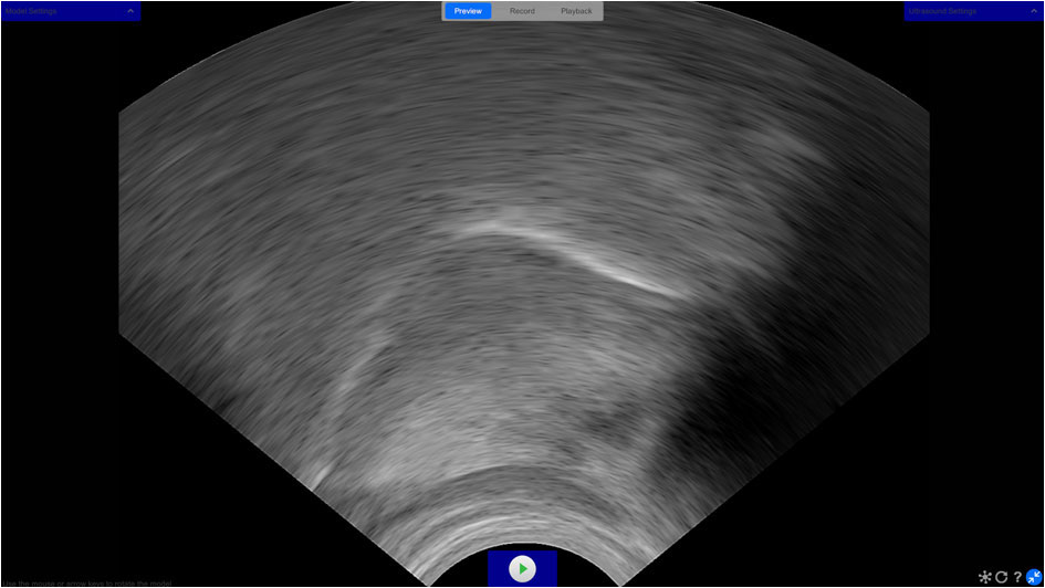
- Real-time 3D tongue, hard palate and teeth visualisation
- Real-time tongue surface contour estimation
- Hard palate estimation
- Probe alignment feedback in Preview, Record and Playback
- 360° photorealistic and stylised views
- Adjustable model transparency
- Synchronised audio recording and playback
- Record at 60+ FPS*
- Preview, record and playback modes
- Real-time export of timestamped tongue contour coordinates and confidence scores to CSV
- Export individual ultrasound video frames as JPG
- Interactive biofeedback game with movable target
- Adjustable targets with touch-activated illumination for biofeedback guidance
- Configurable ultrasound scanner and probe settings
* Test system: Windows 11, AMD Ryzen 7 7700 8-Core Processor (3.80 GHz), NVIDIA GeForce RTX 5060 Ti (8GB) GPU, 16GB RAM
Who uses LinguaSound 3D
LinguaSound 3D can be used by speech professionals, phonetics researchers, pronunciation instructors and human-computer interaction (HCI) engineers.
Clinical
LinguaSound 3D provides speech professionals with a clearer view of tongue position and movement during speech. The 3D model makes it easier to observe articulation patterns and provides real-time visual biofeedback to patients across a range of presentations including speech sound disorders, apraxia of speech and cleft palate. Synchronised audio recording correlates tongue movement with speech sounds, aiding interpretation. Hard palate estimation, which is not possible with conventional ultrasound imaging alone, provides an additional anatomical reference point.
Research
LinguaSound 3D can be used by phoneticians, linguists and speech scientists investigating articulatory phonetics, language-specific tongue gestures and speech production across populations. The software is language independent, making it suitable for research across any language or dialect. Synchronised audio and ultrasound recording supports detailed acoustic and articulatory analysis. Hard palate estimation provides an additional articulatory reference point not available with conventional ultrasound tongue imaging. Real-time tongue surface contour coordinates and timestamps can also be exported for use in custom research and multimodal data collection.
Pronunciation teaching
LinguaSound 3D gives language instructors a precise view of tongue position during speech. Unlike conventional ultrasound tongue imaging, the 3D model requires no specialist knowledge to interpret, making it accessible to instructors and learners alike. It can be used to demonstrate correct tongue placement in real time and provide visual biofeedback during pronunciation practice. Video recordings of tongue movement can also be screen captured directly from the software for use as teaching materials.
Engineering and HCI
LinguaSound 3D outputs real-time tongue surface contour coordinates, confidence scores and timestamps in CSV format at 60+ frames per second, together with the individual ultrasound video frames. This makes it a practical tool for researchers developing assistive technology, silent speech interfaces, and human-computer interaction systems that respond to tongue position and movement.
System requirements
| Parameter | Value |
|---|---|
| Operating system | Windows 10 and 11 (64-bit) |
| Mac support | Not currently supported |
| Supported computers | Desktop and laptop |
| Processor | Intel Core i7 or i9 |
| Graphics | NVIDIA RTX series GPU (8GB) |
| Ram | 16 GB |
| Size on disk | 5 GB |
| Connectivity | USB 2.0 or 3.0 |
| Ultrasound scanner | Telemed MicrUs EXT-1H |
| Ultrasound probes | Telemed MC4-2R20S-3 and MC10-5R10S-3 |
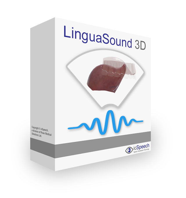
Package contents
LinguaSound 3D is supplied as a perpetual software licence and includes:
- Secure download link to the latest software version
- Software registration key
- Priority technical support
- Free software updates
- 30-day money back guarantee
Note: ultrasound scanner and probe sold separately.
Frequently asked questions
Individual ultrasound images are exported in JPG format. The image size is dependent on the resolution set in the ultrasound settings.
Tongue surface contour data is exported to a CSV file. Eleven labels are used to describe the shape of the tongue surface, along with additional anatomical landmarks including the hyoid bone, mandible base and mental spine. The x, y coordinates of each label are exported relative to the upper left-hand side of the associated image. A confidence score between 0 and 1 is also exported for each label, representing the likelihood that the output of the markerless pose estimation model is correct.
Each row in the CSV file also includes a Time value. In Preview and Record modes, this records the number of milliseconds elapsed since the date and time stored in the file name. Adding the Time value to the file name timestamp gives the date and time at which that row was captured. The time between consecutive rows may vary because rows are written each time new tongue contour data becomes available.
In Playback mode, the Time value represents the position within the original recording and is independent of the date and time stored in the file name. During continuous Playback, a row is written for every video frame, so consecutive rows are evenly spaced according to the recording's frame rate. If a recording is viewed non-sequentially, only the frames actually viewed are exported, so rows may not be evenly spaced.
Data export is supported in Preview, Record and Playback modes.
Ultrasound tongue imaging and electropalatography (EPG) are complementary rather than competing techniques — each captures articulatory information that the other cannot.
Ultrasound shows the shape and movement of the tongue body in real time, including regions that do not contact the palate. It is particularly effective for visualising tongue root position, dorsal gestures, and the full tongue contour in midsagittal view. However, ultrasound cannot directly measure tongue–palate contact.
Electropalatography measures exactly where and when the tongue contacts the hard palate across 62 electrode positions, providing precise, quantifiable contact data. It is particularly effective for sibilant errors, lateral fricatives, and other articulation patterns that midsagittal ultrasound cannot directly capture. Research has shown that lateral fricatives, for example, are easily visualised by EPG but not by midsagittal ultrasound (Cleland et al., 2025).
Both techniques can be used simultaneously using icSpeech Professional Edition, allowing tongue shape and contact patterns to be recorded in a single session.
The software uses the DeepLabCut framework for tongue contour estimation. If you use DeepLabCut as part of your research pipeline, please also cite: Mathis et al. (2018). DeepLabCut: markerless pose estimation of user-defined body parts with deep learning. Nature Neuroscience, 21, 1281–1289.
The tongue contour estimation approach is based on: Wrench, A., and Balch-Tomes, J. (2022). Beyond the Edge: Markerless Pose Estimation of Speech Articulators from Ultrasound and Camera Images Using DeepLabCut. Sensors, 22, 1133.
- CSPNeXt-X — highest accuracy, lowest speed
- CSPNeXt-M — midrange accuracy and speed (default)
- CSPNeXt-S — lowest accuracy, highest speed
Intended use: LinguaSound 3D is speech visualisation software for use by speech and language professionals, researchers, and educators. It is not intended for the diagnosis, prevention, monitoring, or treatment of disease.